
こんちゃです🐸
今回は、前回記事で紹介した「下前腸骨棘下方における関節包付着部の外科的剥離は,術後の関節不安定性の一因になりうる」とする結論が書かれている原著論文について紹介します。
前回記事はこちら↓
そして原著論文はこちら↓

ほんでabstractと翻訳はこちらです↓
原文・翻訳文
題名
An Anatomical Study of the Anterosuperior Capsular Attachment Site on the Acetabulum
寛骨臼の前上方被膜付着部に関する解剖学的研究
Background 背景
Despite the fact that many surgeons perform partial capsular detachment from the anterosuperior aspect of the acetabulum to correct acetabular deformities during hip arthroscopy, few studies have focused on whether these detachments influence hip joint stability. The aim of this study was to investigate the capsular attachment on the anterosuperior aspect of the acetabulum. We hypothesized that the attachment on the inferior aspect of the anterior inferior iliac spine (AIIS) is wide and fibrocartilaginous and might have a substantial role in hip joint stability.
背股関節鏡視下手術において、寛骨臼の変形を矯正するために寛骨臼の前上方から部分的に被膜剥離を行う外科医は多いが、これらの剥離が股関節の安定性に影響するかどうかに焦点を当てた研究はほとんどない。本研究の目的は、寛骨臼の前上方側面における被膜の付着について検討することである。下前腸骨棘(AIIS)下面の付着部は幅が広く、線維軟骨性であり、股関節の安定性に大きく関与している可能性があると仮定した。
Methods 方法
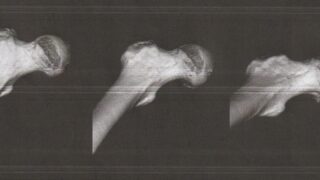
Fifteen hips from 9 cadavers of Japanese donors were analyzed. Eleven hips were analyzed macroscopically, and the other 4 were analyzed histologically. In all specimens, the 3-dimensional morphology of the acetabulum and AIIS was examined using micro-computed tomography (micro-CT).
日本人ドナー9体のうち15体の股関節を分析した.11関節は肉眼的に,残りの4関節は組織学的に解析した.また,すべての標本において、マイクロCT(Micro-Computed Tomography)を用いて寛骨臼とAIISの3次元的な形態を検討した。
Results 結果
Macroscopic analysis showed that the widths of the capsular attachments varied according to the location, and the attachment width on the inferior edge of the AIIS was significantly larger than that on the anterosuperior aspect of the acetabulum. Moreover, the capsular attachment on the inferior edge of the AIIS corresponded with the impression, which was identified by micro-CT. Histological analysis revealed that the hip joint capsule on the inferior edge of the AIIS attached to the acetabulum adjacent to the proximal margin of the labrum. In addition, the hip joint capsule attached to the inferior edge of the AIIS via the fibrocartilage.
マクロ的解析の結果,被膜の幅は部位によって異なり,AIISの下縁の被膜幅は寛骨臼の前上方の被膜幅より有意に大きかった.さらに、AIIS下縁の被膜付着部はmicro-CTで確認した印象と一致した。組織学的解析の結果、AIIS下縁の股関節被膜は臼蓋近位縁に隣接して寛骨臼に付着していることが判明した。また、股関節包は線維軟骨を介してAIIS下縁に付着していた。
Conclusions 結論
The capsular attachment on the inferior edge of the AIIS was characterized by an osseous impression, large attachment width, and distributed fibrocartilage.
AIISの下縁の被膜は,骨性印象,付着幅が大きく,線維軟骨が分布していることが特徴であった。
Clinical relevance 臨床的意義
It appeared that the capsular attachment on the inferior edge of the AIIS was highly adaptive to mechanical stress, on the basis of its osseous impression, attachment width, and histological features. Anatomical knowledge of the capsular attachment on the inferior edge of the AIIS provides a better understanding of the pathological condition of hip joint instability.
骨格印象、付着幅、組織学的特徴から、AIIS下縁の莢膜付着部は機械的ストレスに対して高い適応性を持っているようであった。AIIS下縁の莢膜付着部に関する解剖学的知見は、股関節の不安定性の病態をより良く理解するのに役立つと思われる。
感想
- 共著者に私の執刀医でした先生もおられたので、可及的考慮して手術されたのかが気になるところです。次回受診時に伺ってみたいなと思いました。
- 股関節鏡視下術にも関わる論文の特性上、筆頭著者である理学療法士のTsutsumi PTだけでなく、第一人者であるUchida Dr.と共同研究されたことは術後成績を向上させる可能性のある研究意義がありますね。
- AIIS下縁を一部削るオペで不安定性が可及的軽減できるための考えや対策案は書いて欲しかった。すでにでているのか、対策は最小化アプローチか、それともないのか気になるとこところ。なので論文検索を続けよう🤓


はじめまして。 とても興味をひかれました! ふだん改造ビーチサンダルやlun…
ありがとうございます。嬉しいです みちさんが保存療法でよくなることを願っています…
経過良好で安心しました^_^ やはりリハビリが大事なのですね。 術後の記事、…
お大事に下さい。こちらこそありがとうございました。
コメントありがとうございます。 心強いです。 金曜日に県内のスポーツ外来を受…