
こんちゃ🐸
今回、股関節唇損傷の論文紹介としましたが、股関節包解剖の新たな知見が股関節唇修復術後予後に関わりそうなことを示唆する内容となっています。
解剖学的根拠からリハに役立つ内容と思いますので、興味ある方は是非ご覧ください。
題名
New insight into the iliofemoral ligament based on the anatomical study of the hip joint capsule
股関節包の解剖学的研究に基づく腸大腿靭帯に関する新しい知見

Abstract
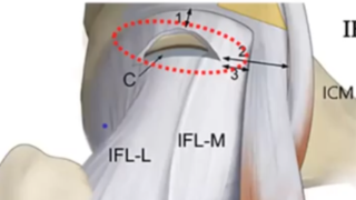
The iliofemoral ligament, which plays an important role in hip joint stability, is formed on the anterosuperior region of the hip joint capsule.
股関節の安定性に重要な役割を果たす腸大腿靭帯は、股関節包の前上方部に形成されている。
Although the tendon and deep aponeurosis of the gluteus minimus and iliopsoas are partly connected to the same region of the capsule, the precise location of the connections between the joint capsule and the tendons and deep aponeuroses remains unclear.
小殿筋と腸腰筋の腱や深部骨膜は、関節包の同じ部位に一部つながっているが、関節包と腱や深部骨膜の正確な接続位置は不明なままである。
The locations of the tendinous and aponeurotic connections with the joint capsule may clarify whether the iliofemoral ligament can be regarded as the dynamic stabilizer.
腱や骨膜と関節包の結合部位から、腸骨大腿靭帯が動的安定化因子と見なせるかどうかが明らかになるであろう。
This study investigated the relationships between the anterosuperior region of the joint capsule and the tendon and deep aponeurosis of the gluteus minimus and iliopsoas. Fourteen hips from nine cadavers (five males; four females; mean age at death 76.7 years) were analyzed.
本研究では、関節包の前上方領域と小殿筋および腸腰筋の腱および深部骨膜との関係について検討した。9体の死体から得られた14個の股関節(男性5人、女性4人、死亡時平均年齢76.7歳)を分析した。
Ten hips were macroscopically analyzed, and four were histologically analyzed. During macroscopic analysis, the joint capsule was detached from the acetabular margin and the femur, and its local thickness was measured using microcomputed tomography (micro-CT).
10個の股関節が肉眼的に分析され、4個が組織学的に分析された。巨視的解析では、関節包を臼蓋縁と大腿骨から剥離し、その局所的な厚みをマイクロCT(micro-computed tomography)で測定した。
The gluteus minimus tendon was connected to the joint capsule, and the lateral end of this connection was adjoined with the tubercle of the femur at the superolateral end of the intertrochanteric line.
小殿筋腱は関節包に接続され,その外側端は大腿骨転子間線の上外側端で大腿骨の結節と隣接していた.
he deep aponeurosis of the iliopsoas was also connected to the joint capsule, and the inferomedial end of its anterior border corresponded with the inferomedial end of the intertrochanteric line.
腸腰筋の深部筋膜も関節包に連結しており、その前縁の内側端は転子間線の内側端に対応していた。
In the micro-CT analysis, capsular thickening was observed at the base of the connection to the gluteus minimus tendon and at the anterior border of the deep aponeurosis of the iliopsoas.
マイクロCT解析では、小殿筋腱との接続基部と腸骨深層筋の前縁に被膜の肥厚が観察された。
A histological study showed that the gluteus minimus tendon and the deep aponeurosis of the iliopsoas were continuous with the hip joint capsule.
組織学的な検討の結果、小殿筋腱と腸腰筋の深部骨膜は股関節包と連続していることがわかりました。
Based on the morphology of the tendinous and aponeurotic connections, local capsular thickening and histological continuity, the transverse and descending parts of the iliofemoral ligament were the joint capsules, with fibers arranged according to the connection with the gluteus minimus tendon and the deep aponeurosis of the iliopsoas, respectively.
腱・腱膜の接続形態、局所的な被膜の肥厚、組織学的連続性から、腸大腿靭帯の横方向と下方向の部分が関節被膜であり、それぞれ小殿筋腱と腸腰筋の深部被膜との接続に従って繊維が配置されていた。
Therefore, the so-called iliofemoral ligament could be regarded as the dynamic stabilizer, with the ability to transmit the muscular power to the joint via the capsular complex. This anatomical knowledge provides a better understanding of the hip stabilization mechanism.
したがって、いわゆる腸骨大腿靭帯は、莢膜複合体を介して関節に筋力を伝達する機能を持つ、動的安定化装置とみなすことができる。このような解剖学的な知見は、股関節の安定化機構をより深く理解するためのものです。
Keywords: gluteus minimus; hip joint capsule; iliofemoral ligament; iliopsoas.
キーワード:小殿筋、股関節包、腸大腿靭帯、腸腰筋。
© 2019 The Authors. Journal of Anatomy published by John Wiley & Sons Ltd on behalf of Anatomical Society.


はじめまして。 とても興味をひかれました! ふだん改造ビーチサンダルやlun…
ありがとうございます。嬉しいです みちさんが保存療法でよくなることを願っています…
経過良好で安心しました^_^ やはりリハビリが大事なのですね。 術後の記事、…
お大事に下さい。こちらこそありがとうございました。
コメントありがとうございます。 心強いです。 金曜日に県内のスポーツ外来を受…